Pollen Data Table
Friday, December 20, 2013
Monday, December 16, 2013
Procedure
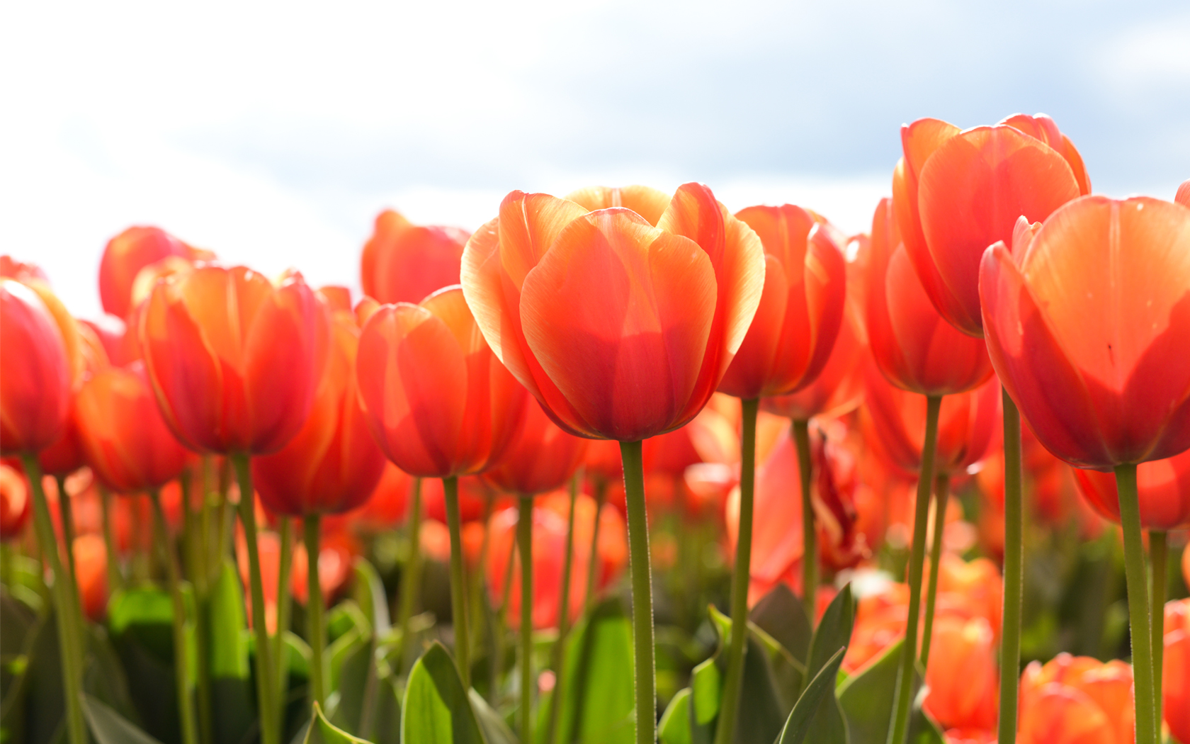
First we gathered red roses (Rosa berberifolia) from Betty's garden, tulips (Tulipa gesneriana) from Mrs. Lindahl's collection, and winter pansies (Viola tricolor) from Mrs. Lindahl's collection. To prepare the roses, we put them in the dehyrator for 48 hours to dry them out because the SEM cannot have any moisture inside of it. The tulips and pansies were already dried out.

Rose


Winter Pansy

Step Two:
Then we loaded the stub that we'll use in the SEM microscope. We took the stub and peeled off the white tape. We pushed the stub on to a piece of carbon tape that was on a sheet, then peeled the stub (attached to the carbon tape) off of the carbon shape.
We gathered all of our samples--the roses, tulips, and winter pansies. For each sample we had a different paintbrush. One at a time, we used the appropriate paintbrush to brush a sample of pollen onto the stub.There were three different sections on the stub, one for each of the pollen samples.
When we had finished loading the pollen samples on the stub, we blew off the stub with a multi-purpose duster, so that loose pollen wouldn't come off in the SEM machine. Then the stub samples were prepared, so we put the stub into a small white box labeled with our names for storage.
Step Three:
We prepared pollen samples to look at under the microscope. We took brushes and brushed pollen from each flower onto a glass slide. Then we added a drop of water, and put a cover slip over it. Under a Swift Ultra microscope, we looked at each sample under 400x. Unfortunately, we couldn't find any pollen from the winter pansy, but we got photos of the rose's and tulip's pollen.

Tulip pollen, 400x

Rose pollen, 400x
Step Four:
Next we looked at our samples under a Leica EZ4 HD microscope, using Leica Imaging 1.0. We looked at our prepared stub, and our original flower collections that were in bags. We looked at the winter pansy, tulips and roses one at a time under the microscope. Unfortunately, our Leica photos were never loaded off of the computer before the computer was taken away, so we don't have those photos. However, we have comparable photos taken on a Frey Scientific microscope at 35x.

Rose, 35x

Red tulip, 35x

Winter pansy, 35x
Step Five:
Now we used the SEM (Scanning Electron Microscope) to take pictures of our samples. First, we picked up the stub with two fingers from the storage box. We took the cup from the SEM, and pushed the stub down into the cup. We lowered the sample to be even with the cup (we eyed it), by turning the silver top part. Then we used the side notch on the cup, and clicked over four times to the right, which makes sure that the sample is low enough to go into the SEM. We placed the cup into the slot of the SEM, and pushed all the way in, until the green light came on. Then we pushed down the door of the SEM.
On the screen, we pressed the scanning button. We mapped the entire sample by pressing the button that looked like a maze. Then we went into settings and pressed label to label our sample, then clicked okay, then went back to the image. We started scanning our sample, and when it was ready we moved around it and took pictures. Here are the pictures we took below.
Tulip Pollen Pictures

Collection of several pollen grains, 360x.

Pollen grain, 2720x.

Texture of pollen grain, 5050x

Pollen grain, including the length measurement, 2000x.
Rose Pollen Pictures

Collection of pollen grains, 2600x.

Grain of pollen, 4200x.

Grain of pollen that shows its three apertures, 6500x.

Same grain of pollen as above, 12000x.

Grain of pollen that shows the measurement of the length of the grain (25.6 um)
2000x.

Same pollen grain as above, displays the length and width measurement of the grain (45 um long) 2000x
Winter Pansy Pictures

One grain of pollen, showing an aperture, 725x
Photo from http://pollenlab.blogspot.com


Pollen grain with decollate apertures, shows length measurement of grain (75 um), 2000x
Photo from http://pollenlab.blogspot.com
Photo from http://pollenlab.blogspot.com


Step Six:
The first thing that we did was we looked at all the background that we currently had and we decided that what the two closes relating flowers would have to be. With that information we made our pollen morphology.
Step Seven:
What we did was find out what the names of our flowers were and we went on a web sit that gave us information about our flowers. Then we found out which flowers were really related because of what flowers had the most proteins they had in common. Finally we made a tree of our flowers by protein sequence.
Hypothesis
We think the pollen in winter flowers would have more of a complex structure because it's harder to bloom in the winter due to the weather and climate.
Background
We looked at this article which said that cold temperatures can affect the pollen of plants. Cold temperatures are needed for many plants to bloom.
http://www.desertmuseum.org/programs/flw_predicting.php
This article said that when plants have less water during flowering time, the pollen is larger and more spherically shaped. We thought this might apply to winter flowering plants because there is less rainfall during winter than there is during spring.
http://www.academia.edu/2393597/Does_climate_affect_pollen_morphology_Optimal_size_and_shape_of_pollen_grains_under_various_desiccation_intensity
This article said that the pollen of spring pollen was different in form and size from summer flowering pollen.
http://www.doiserbia.nb.rs/img/doi/0354-4664/2012/0354-46641201085B.pdf
Question
What is the difference in pollen in flowers that bloom in spring vs. flowers that bloom in the winter?
Team Agreement
Vivy Kaye-Diamond: Researched which pollens to use, researched background info, Step One of procedure, Step Two of procedure
Shawna Blazer:
Lily Gould: Step One of procedure
Betty Glanz: Brought in rose pollen, loaded all pollens onto stub, researched background info, recorder while using SEM microscope
Addendum 12-20-13
Vivy Kaye-Diamond: Working on addendum, typing up our findings of background research, finishing step two and three of procedure.
Shawna Blazer: looking up internet photos for procedure
Lily Gould: filling out Pollen Data Table and attaching it to blog
Betty Glanz: searching for photos on flashdrive
Adendeum Winter Break
Vivy Kaye-Diamond: searching for more background information
Shawna Blazer: Highlight SEM photos
Betty Glanz: uploading SEM photos to procedure section, determine what genus of rose we're using by looking at SEM photos
Adendum 1-6-14
Vivy Kaye-Diamond: Started writing descriptions for SEM photos
Shawna Blazer: Started pollen morphology tree
Lily Gould: Started Pollen Data Table
Betty Glanz: Uploaded SEM photos to Procedure
Adendum 1-8-14
Vivy Kaye-Diamond: Moved pictures from SEM into appropriate section in Procedure, wrote descriptions of each picture, updated Team Agreement.
Shawna Blazer: Created protein sequence tree, took screenshot of the tree, made pollen morphology tree
Lily Gould: Finished Pollen Data Table
Betty Glanz: Looked for SEM winter pansy photos to borrow and cite, uploaded borrowed pansy photos, emailed other team to ask if it was okay to borrow pansy photos
Adendum 1-10-14
Vivy Kaye-Diamond: Updated team agreement, worked on SEM step of the procedure (rewatched video)
Shawna Blazer: Uploaded both trees to blog, worked on analysis of trees
Lily Gould: Absent
Betty Glanz: Worked on analysis of question and how data can be interpreted
Shawna Blazer:
Lily Gould: Step One of procedure
Betty Glanz: Brought in rose pollen, loaded all pollens onto stub, researched background info, recorder while using SEM microscope
Addendum 12-20-13
Vivy Kaye-Diamond: Working on addendum, typing up our findings of background research, finishing step two and three of procedure.
Shawna Blazer: looking up internet photos for procedure
Lily Gould: filling out Pollen Data Table and attaching it to blog
Betty Glanz: searching for photos on flashdrive
Adendeum Winter Break
Vivy Kaye-Diamond: searching for more background information
Shawna Blazer: Highlight SEM photos
Betty Glanz: uploading SEM photos to procedure section, determine what genus of rose we're using by looking at SEM photos
Lily Gould: analyze SEM photos and fill out Pollen Data Table
Vivy Kaye-Diamond: Started writing descriptions for SEM photos
Shawna Blazer: Started pollen morphology tree
Lily Gould: Started Pollen Data Table
Betty Glanz: Uploaded SEM photos to Procedure
Adendum 1-8-14
Vivy Kaye-Diamond: Moved pictures from SEM into appropriate section in Procedure, wrote descriptions of each picture, updated Team Agreement.
Shawna Blazer: Created protein sequence tree, took screenshot of the tree, made pollen morphology tree
Lily Gould: Finished Pollen Data Table
Betty Glanz: Looked for SEM winter pansy photos to borrow and cite, uploaded borrowed pansy photos, emailed other team to ask if it was okay to borrow pansy photos
Adendum 1-10-14
Vivy Kaye-Diamond: Updated team agreement, worked on SEM step of the procedure (rewatched video)
Shawna Blazer: Uploaded both trees to blog, worked on analysis of trees
Lily Gould: Absent
Betty Glanz: Worked on analysis of question and how data can be interpreted
Subscribe to:
Comments (Atom)